SCIENCE OF
THE BLUE WALLEYE OF CANADA by Dr. Wayne Schaefer
As a fish
biologist and university professor, I had worked with walleye in Ontario for
nearly 10 years but had never seen a blue one.
While a student, I was taught the importance of blue pike (really a walleye)
in the history of Lake Erie where commercial catch of that species was a major
part of the market in the early 1900s.
However, the species had been driven to extinction by over-fishing and
pollution by the mid-1950s and no one had seen one since that time. Therefore, I was greatly surprised to see a
blue colored walleye on the end of my line while fishing the Papaonga River
System in Northwestern Ontario. The riiver
is near my summer cabin on Pakwash Lake near Ear Falls (Figure 1).

It was a warm August day in 1993 when we slipped a canoe into McKim Lake. The lake seemed perfect for a canoe. It was small (only 2 miles across) with a medium sized river going through its eastern basin and with easy access from a gravel logging road just 35 miles east of Ear Falls. We worked the canoe to the east side of the lake where the river entered and started to catch walleye that had a beautiful turquoise blue color on their dorsal (upper) and caudal (tail) fins (Figure2).

Closer examination of the fish showed the blue color to be present only on the dorsal part of the fish, above the lateral line (Figure 3).

The belly of the fish was white with no
yellow pigmentation. Could this be a
remnant population of blue pike?

It was a warm August day in 1993 when we slipped a canoe into McKim Lake. The lake seemed perfect for a canoe. It was small (only 2 miles across) with a medium sized river going through its eastern basin and with easy access from a gravel logging road just 35 miles east of Ear Falls. We worked the canoe to the east side of the lake where the river entered and started to catch walleye that had a beautiful turquoise blue color on their dorsal (upper) and caudal (tail) fins (Figure2).

Closer examination of the fish showed the blue color to be present only on the dorsal part of the fish, above the lateral line (Figure 3).

The next
spring I started asking Canadian outfitters at sport shows in Wisconsin (my
home state), Illinois and Minnesota if they had any blue walleye in their
lakes. Most outfitters responded with
“There is no such thing as a blue walleye”.
However, the guys at Wilderness North Outfitters said they had blue
walleye in a small lake out of Armstrong, Ontario, just north of Lake
Nipigon. I made arrangements to fly into Dawn Lake the
next May. The single cabin on the lake
was well cared for and the boats and motors were in good shape. It wasn’t difficult to locate walleye but
none were blue in color. Instead they
were unusually gray in color with very little, if any, yellow coloration. They tasted just as good as any walleye I had
ever eaten. Out of desperation, I put a
couple whole fish into the cooler in zip lock bags, with a little lake water,
and brought them back to my lab at the University of Wisconsin Milwaukee –
Washington County. To my surprise, upon
opening the cooler a couple days later, the water in the bags was colored
bright blue. The color could only have
come from the mucus of the fish. After
searching the scientific literature, I found no other report of color in the
mucus of any fish. The most logical
explanation was a symbiotic micro-organism in the skin mucus of the fish. Surely it would be easy to isolate the
organism and produce blue color on a nutrient substrate. I enlisted the help of many students, and a
few colleagues, in an effort to isolate the suspected bacterium or algae that must
be producing the blue color, but we were unable to identify any such
micro-organism that produced blue color on any type of nutrient agar, and we
tried them all. About that time, I took
on a Ph.D. student, Mark Schmitz, who was enrolled at the University of
Wisconsin – Milwaukee. I was his
co-advisor and his dissertation would be “the biology of blue walleye in
Canada”.
After
talking to several well-known microbiologists at big universities we learned of
a professor at the University of Iowa who was working on blue color in
bacteria. His name was Dr. David Gibson
and he was a world renowned scientist.
With a little bit of timidity, I emailed the expert and, to my surprise,
he replied, “I have two passions in life – microbiology and fishing”. That was the start of one of the most
fulfilling collaborations of my life.
Dr. Gibson put a bright young female postdoc student on the
project. Her name was Chi-Li Yu. Since Chi-Li was from China, she had never
seen a walleye before in her life but she did know protein chemistry. Within two months Chi-Li had isolated the
blue pigment and determined it to be a medium-sized protein, secreted by the fish,
with molecular weight 87,850 Daltons.
The protein was a tetramer, consisting of 4 identical sub units and belonged
to a family of proteins called lipocalins which were known to act as carriers
of specific smaller molecules referred to as ligands. A couple months later Chi-Li and David had
identified the ligand as biliverdin, a common excretory product in vertebrate
animals. We named this previously
unknown protein, “sandercyanin”. Sander
is the genus name for walleye and cyanin means blue in Greek. Sounded pretty scientific. We published that early work in 2008 in the
Journal of Fish Biology (82(1): 51-58). After David’s retirement in 2004, Dr. Ramaswamy
Subramanian (Rams), continued working with Chi-Li in Iowa until he transferred
to India to work as a supervisor in the world renowned Institute for Stem Cell
Science and Regenerative Medicine (inStem) in Bangalore. He, and one of his doctoral students,
Swagatha Ghosh, continued to work on the chemistry of sandercyanin and
accomplished remarkable things, which we will discuss later in this article.
The answer
to the most important question still had eluded us, “Could these fish represent
a remnant population of the extinct blue pike of Lake Erie”? Maybe we could restock Lake Erie and bring
back this important species. The only
true test would be genetic analysis.
Fortunately, an expert in walleye genetics was only a couple hundred
miles away from my home campus and taught at the University of Toledo. Dr. Carol Stepien was excited to see the
fish. Not only had she worked on
present-day walleye but had also done genetic analysis on museum specimens of
extinct blue pike housed at the University of Michigan. If anyone could get the answer, it would be
Dr. Stepien. After careful analysis of
the DNA from our Canadian walleye, Dr. Stepien determined that the fish were
not the same subspecies (glaucus) as
the extinct blue pike of Lake Erie. In
fact they were simply a color variant of regular, run of the mill, walleye
(Sander vitreus). That shot down our
second hypothesis, the first one being the expected presence of a blue
symbiotic micro-organism in the mucus of the fish. We had, however, determined two possible reasons
why the fish were blue in color. First,
they were albino for yellow color in their skin and, second, they produced a
blue pigment in their skin mucus that was particularly evident on the dorsal
side of the fish. Also, the production
of blue pigment (sandercyanin) seemed to be seasonal, peaking in late
summer.
We decided
to document intensity of blue color in the mucus of the fish over all four
seasons, which was particularly challenging in winter. We used my cabin on Pakwash Lake as a base
camp and traveled 50 miles, one way, to McKim Lake each day, sometimes through
snow and ice or mud, and in the summer, always with mosquitos and black flies, and
sometimes bears, on gravel logging roads to get specimens. However, I always did say that the best part
of this project was our method of sampling fish – hook and line! Not only that but, unlike most researchers in
biology, we got to eat our specimens!
Blue walleye taste just as good as regular walleye! Another interesting observation was that the
blue color does not come off the fish onto your hands when you handle them,
however, it does come off onto a knife blade when you scrape the dorsal (upper)
side of the fish from head to tail (Figure 4).

The blue color
is readily apparent against a white background of snow (Figure 5) and is also seen in snow around
the fish when you are ice fishing.

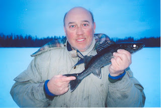
The
author is shown in Figure
6 holding a blue walleye on the ice of McKim Lake.
Eventually we did obtain samples of walleye
mucus from over 300 fish across all four seasons during a period of six years
and, with the help of other researchers from the University of Wisconsin Milwaukee,
tested those samples for seasonal intensity of blue color. The data turned out perfect (Figure 7).

The intensity of blue color in the mucus
peaked dramatically each year in late summer.
The pieces of the puzzle, as to why Canadian walleye produce
sandercyanin, were beginning to fall into place.


In an effort
to determine exactly where on the fish sandercyanin was being produced we
examined fresh specimens of blue walleye under the microscope. In Figure 8 is shown a close up of the spines in the anterior
dorsal fin.

Notice the blue line behind
each spine which runs parallel to a blood vessel in each spine. Clearly sandercyanin was being produced just
behind each dorsal spine in the walleye fin. As noted previously, blue color was only
produced above the lateral line. Closer
examination of the blue color showed that sandercyanin was packaged in cellular
vesicles in the mucus of the fish (Figure 9).

Blue walleye
are the only fish in the world, so far as we know, that produce color in their
skin mucus. Further, no other fish
species, that we could find, packages pigment in cellular vesicles. These results caused us to make histological
cross sections of the skin and examine them under a microscope at higher
magnification (Figure 10).

Dr.Vicki Blazer at the National Center
for Fish Disease in Kearneysville, West Virginia was gracious enough to help us
with that project. In Figure 10, the area above
the surface of the skin appears white in the upper left. The blue ovals represent the cells producing
sandercyanin. The white spaces, just
below the surface of the skin, are the locations of empty mucus-producing cells. The cells producing sandercyanin are just
below the mucus cells and are blue in color.
They are a type of “sacciform” cell.
Sacciform cells occur rarely in fish species and generally store
important chemicals needed by the surrounding tissue. We have found no other report in the
literature of a sacciform cell that stores pigment.


From the
start of this research, I had wondered if the blue color of sandercyanin might
fluoresce in ultraviolet (UV) light. All
attempts we made to “light up” whole specimens of blue walleye in UV light
failed. However, in June of 2008, while working
in the microscope lab at the University of Iowa, we radiated samples of blue
mucus, from fresh Canadian walleye, with UV light under the microscope. To our amazement, the cellular vesicles
containing sandercyanin lit up red like a Christmas tree. Sandercyanin was a fluorescent protein (Figure 11)!

Several of these remarkable proteins have
been found in simple animals around the world, including the Nobel Prize winning,
and very useful, green fluorescent protein from jelly fish. Who would have guessed that walleye in Canada
might produce a fluorescent protein?
Rams and Swagatha cloned the gene for sandercyanin and we patented it
through the University of Wisconsin. The
gene has been transferred to bacteria which now produce the blue colored
Sandercyanin. However, to our great
disappointment, the recombinant sandercyanin does not fluoresce well in UV
light like the native protein does in vesicles.
Rams and his colleagues in India are attempting to engineer the recombinant
protein to fluoresce brighter but, so far, have been unsuccessful.

Another big
question loomed over our research. I had
established a blog website (bluewalleye.com) where sport fishermen could report
the geographic location of blue walleye.
Hundreds of sightings came into the site from across Ontario and Quebec
but very few from south of the Canadian border.
Why were blue walleye mostly found in the northern part of the range of
walleye, above the Canadian border, and only rarely in the United States, and
did that observation go along with the observations that sandercyanin was
produced more in the summer and only on the dorsal part of the fish? The answer to that question came from a high
school student, one of over three hundred, who helped us with chemical analysis
of the blue mucus. He simply stated “Dr.
Schaefer, maybe it is related to the ozone hole over the North Pole”. As soon as he said it, I knew it was
true. It was the most reasonable
hypothesis to tie together all three of our major observations – sandercyanin
was only produced on the dorsal side of the fish where the sun hits it. It was produced seasonally during the period
of longest day-length and it was produced only in the northern part of the
hemisphere where an ozone hole existed in the earth’s atmosphere. Ozone there is destroyed by chlorofluorocarbon
(CFC), an air pollutant. Ozone, which naturally
exists in the upper atmosphere of the earth, absorbs harmful UV radiation from
the sun, thereby protecting life on earth.
With an ozone hole over the North Pole, walleye in that area would be
exposed to higher levels of harmful UV radiation than their cousins to the
south.
How did UV
radiation cause increased production of sandercyanin? The answer to that important question is in
the fact that UV radiation causes the breakdown of heme, the red protein of
blood, into biliverdin, the ligand carried by sandercyanin. Excess biliverdin is excreted from the skin
of the fish and combines with the protein Sandercyanin causing it to turn blue
in color. Further, analysis of the
photo-absorption spectrum of Sandercyanin revealed that its highest absorption
of light was in the UV range (300-400 nanometers), thereby protecting deep skin
tissue from the harmful effects of UV light.
It was all starting to come together.
Walleye in the upper part of North America were producing sandercyanin
to protect themselves from exposure to high levels of UV light. Ironically, the fish were using the very
product (biliverdin), resulting from damage to their skin by exposure to UV
radiation, to protect them from the cause of that damage (UV radiation). This is a beautiful story of nature
protecting itself from human interference.
Perhaps,
this is how science is done. You ask
questions and follow the answers until the truth reveals itself. Most science seems simple, once you know the
answers. The answers to these questions
came only by diligent research from many tough students and many tough
colleagues, to which I am most grateful.
It has been my joy to work on blue walleye for over 16 years, if you can
really call “fishing” work! We were
privileged to work in some of the most beautiful wilderness country in the
world (Figure 12)
and were treated many times to northern lights at night (Figure 13).


It has been said that if you enjoy your work, you will never work a day of your life. Such has been my happy lot. It is my hope that this article will instill within walleye fisherpersons even a greater appreciation for this beautiful fish.


It has been said that if you enjoy your work, you will never work a day of your life. Such has been my happy lot. It is my hope that this article will instill within walleye fisherpersons even a greater appreciation for this beautiful fish.
To those who
would like more detailed information on the science of blue walleye, please
check out the links below to our two most recent publications:
Schaefer, Wayne F., Schmitz, Mark H., Blazer, Vicki S.,
Ehlinger, Timothy J., Berges, John A.
2015. Localization and
seasonal variation of blue pigment (sandercyanin) in walleye (Sander
vitreus). Canadian Journal of Fisheries
and Aquatic Sciences 72(2): 281-289.
Ghosh, S.; Yu, C.; Ferraro, D.J.;
Sudha, S.; Samir, K.P.; Schaefer, W.F.; Gibson, D.T.; Ramaswamy, S. 2016. A Blue Protein with Red Fluorescence. Proceedings of the National Academy of Sciences of the United
States of America, 113(41):11513-11518.
To those who
would like to catch a blue walleye, just contact the outfitters below and they
will set you up in a remote camp in the wilderness of Canada where blue walleye
are present.
Excellent
Adventures in Ear Falls, Ontario.
Exc-adventures.com. 807-662-5292.
Northern Wilderness
Outfitters in Thunder Bay, Ontario.
Wildernessnorth.com.
888-465-3474.
Ghost River
Outfitters in Sioux Lookout, Ontario.
Ghostriverlodges.com.
888-446-7874.
Next time
you catch a walleye, scape the skin from head to tail on the dorsal side of the
fish with a knife and see if blue color is present. If so, please report the finding on my
website at bluewalleye.com. Good
fishing!


